Author : Charles J. Goodacre
Edition : 2nd Edition
Download PDF Atlas of the Human Dentition
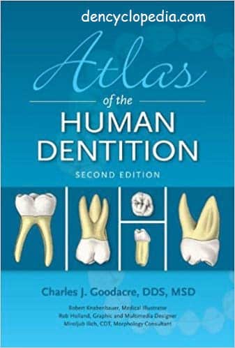
This atlas has been very beneficial for dental students, dental auxiliary college students, dental laboratory technicians, and every body interested by gaining knowledge of or reviewing the specific morphologic characteristics of human tooth.The drawings consist of all of the primary and secondary enamel from five perspectives (facial, lingual, mesial, distal, and incisal/occlusal). in addition, there are unique drawings that allow clean visible comparisons to be made between the one-of-a-kind primary tooth, the one-of-a-kind secondary teeth, and additionally comparisons to be made among the primary and secondary teeth.
This Atlas consists of drawings of each number one and secondary enamel from five perspectives (facial, lingual, mesial, distal, and incisal or occlusal) and composite drawings of multiple tooth that permit visual morphologic comparisons to be made. The tooth had been drawn so they mirror the traits defined with the aid of the author. The drawings in this atlas consist of all the primary and secondary teeth from 5 perspectives (facial, lingual, mesial, distal, and incisal/occlusal). in addition, there are specific drawings that permit clean visible comparisons to be made among the distinctive primary enamel, the exclusive secondary teeth, and additionally comparisons to be made between the number one and secondary teeth.
The Atlas of the Human Dentition designed for smooth viewing of the 77 pages of particular drawings. all the drawings are scale drawings, based at the “average” dimensions of each enamel as recognized inside the dental literature. additionally, a completely unique characteristic is that every one of the key morphologic functions typically observed of every enamel are seen at the drawings. The Atlas additionally includes tables that display the common dimensions as decided in the dental literature, in addition to tables that identify the coronal outline shape, coronal heights of contour, marginal ridge locations and bureaucracy, root floor and morphology, proximal contact paperwork and region, and cervical crown form.


